0
Skip to Content
The Roberson Lab
Lab Members
Lab News
Publications
Collaborators
Contact us!
The Roberson Lab
Lab Members
Lab News
Publications
Collaborators
Contact us!
Lab Members
Lab News
Publications
Collaborators
Contact us!
2/29/24

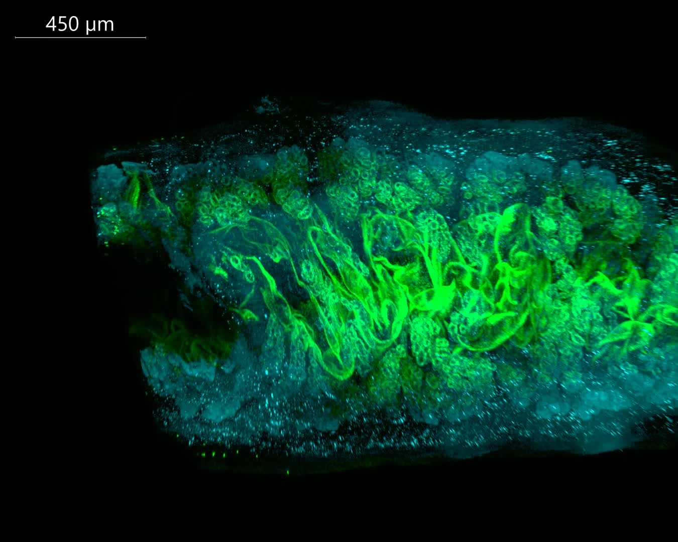
3D Segmentation of Uterine Glands from Volumetric Images

Wild type mouse in in diestrus. E-cadherin (epithelial cell adhesion marker) shown in cyan. Foxa2 (gland determining transcription factor) depicted in magenta. Glands are individually pseudo colored for easy visualization.

Next

Immunofluorescent Volumetric Imaging of all Epithelium

You Might Also Like

Related Video Item Thumbnail Immunofluorescent Volumetric Imaging of all Epithelium