0
Skip to Content
The Roberson Lab
Lab Members
Lab News
Publications
Collaborators
Contact us!
The Roberson Lab
Lab Members
Lab News
Publications
Collaborators
Contact us!
Lab Members
Lab News
Publications
Collaborators
Contact us!
2/29/24

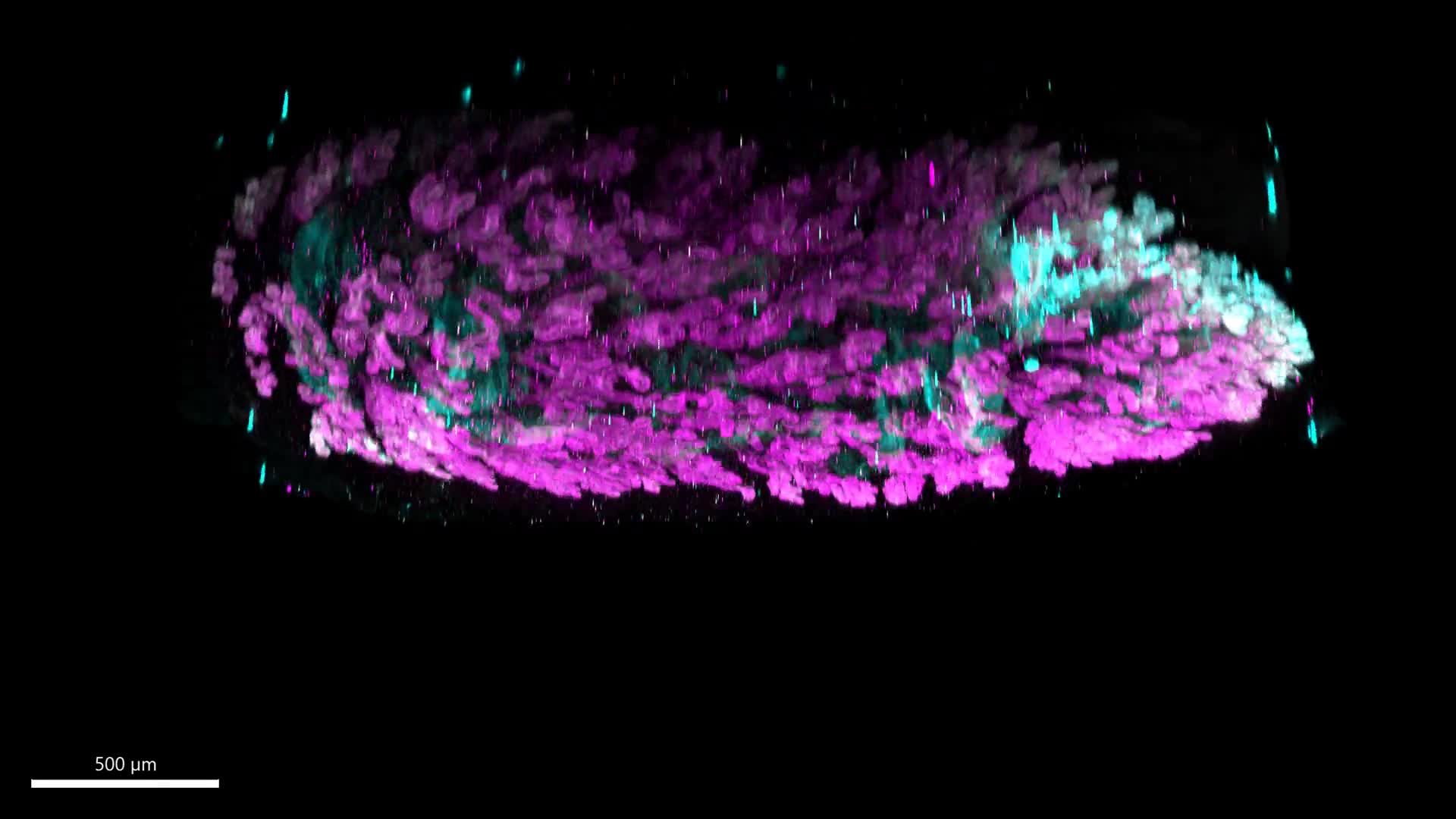
Immunofluorescent Volumetric Imaging of all Epithelium

Wild type mouse in metestrus. GFP (endogenously expressed in epithelial cells, driven by Pax8 expression) is depicted in green in all epithelia. E-cadherin (epithelial cell adhesion marker) is shown in cyan.

Previous

3D Segmentation of Uterine Glands from Volumetric Images

You Might Also Like

Related Video Item Thumbnail 3D Segmentation of Uterine Glands from Volumetric Images